What Is Limiting Your Ankle Mobility?
Ah, ankle joint dorsiflexion. One of the most talked about joints when it comes to joint mobility and optimal movement patterns.
Tight soleus, tight gastrocnemius, heel toe drop in shoes, flat feet, high arches......sometimes it seems overwhelming to know exactly what is the cause of this restricted mobility in our clients.
This article will review some key concepts when it comes to ankle joint mobility and how to address one of the most overlooked causes of restricted dorsiflexion - the anteriorly shifted talus!
Why do I even need ankle joint dorsiflexion?
Joint mobility, particularly in the foot and ankle, is necessary for proper loading and unloading of impact forces and in transferring the associated potential energy.
When our joints move, in this case the ankle, there is a stretching (or potentiating) of the Achilles tendon and surrounding fascial structures. You can think of this like stretching a rubber band or pulling the string back on a bow and arrow.
The stretching of a rubber band or bowstring represents the potential energy that we will then release and use to catapult our leg forward when we take the successive step.
If that joint mobility is restricted then there is less potentiation or stored energy in the tendon, and instead of recoiling our leg forward we pull our leg forward when we walk.
This can lead to an increased work load, accelerated musculo-tendinous fatigue and increased injury risk.
So how much dorsiflexion do we need, and how do we assess for it?
Well, it depends. It depends on what you are doing. Are you concerned about ankle joint dorsiflexion to squat? Or ankle joint dorsiflexion to walk?
When I assess a patient I am primarily concerned about their ankle joint dorsiflexion when walking, as this is the pattern we do 10,000+ times a day. I often say that if we cannot do the baseline functional movement demands of walking then we shouldn't be deviating into more challenging movement patterns.
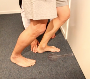
If you are concerned about ankle joint dorsiflexion for walking - this assessment must be done in a straight leg position as that's how our ankle dorsiflexes when walking.
Most of the videos and articles I see on the internet demonstrate a knee-bent ankle joint assessment technique such as that pictured to the right. Now this assessment technique is not wrong - it just doesn't apply to walking.
My preferred open chain assessment method would have the client lying on their back and then the subtalar joint is put into neutral and ankle dorsiflexed maximally.
I like to look for at least 5 degrees of dorsiflexion in this position.
This assessment can be followed by a Silverskold test which is where the client would bend their knee to determine if there is an increase in range of motion caused by a tight gastrocnemius muscle.
Now you may be wondering - why is the STJ in neutral during this assessment?
And why do we need only 5 degrees of ankle dorsiflexion to walk?
That seems like very little dorsiflexion!
When we walk the point in gait where we need maximum ankle dorsiflexion is in late midstance, or when our leg is behind us and the heel is down.
In this position - in this moment in time - in this few milliseconds - we are simply passing through our ankle on our way to becoming a rigid lever that is supinated and stable for push off. This means that we do not want to fully pronate as we go into ankle joint dorsiflexion. By keeping the STJ in neutral this restricts ankle dorsiflexion and helps to maintain a stable foot for ambulation. (Still confused on this concept then please do check out our full training and Certification\ Barefoot Training Specialist Level 1)
The closed chain assessment I do for the ankle joint dorsiflexion is a gait assessment. By simply watching how a client goes through late midstance it will tell me if they have sufficient functional ankle joint dorsiflexion.
What happens if we don't have 5 degrees of dorsiflexion when walking?
We compensate. Period.
Your ankle and foot will find a path of least resistance and find a way to get through late midstance. You could turn your foot out and walk like a duck. You could pronate as you go through late midstance. You could lift your heel early, creating a bouncy gait we see in some of our friends.
Or simply stated, your body will find a way to get through late midstance that is less efficient, stores less potential energy and increases your risk of soft tissue or joint injury.
This is why it's important to not only know how to assess for proper ankle joint dorsiflexion - but also what is causing it and how to fix it!
So how do we increase ankle joint dorsiflexion?
As I said above. You need to know why is the ankle restricted in the first place?
Many professionals will only, or primarily, think of soft tissue reasons for limited ankle dorsiflexion. This includes tight soleus, tight gastrocs, tight calves, tight Achilles tendon, tight plantar fascia - however the causes of ankle joint restriction are not limited to soft tissue.
One of the most overlooked causes of limited ankle dorsiflexion is - the boney block!
What is a boney block?
There are a couple reasons why clients can get a boney block at the ankle joint with the most common being spurs / osteophytes or an anteriorly shifted talus. For the sake of this article we are going to focus on the anteriorly shifted talus.
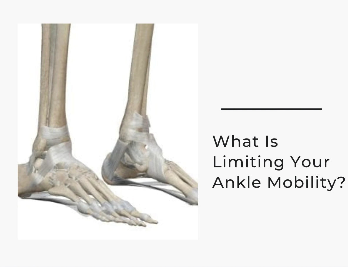
Meet the talus. This bone is a fascinating bone in that it articulates with three joints - the ankle, the subtalar joint, and the talonavicular joint. It also is kind of like a floating bone in that it has no muscle or tendon attachments!
To repeat. The talus is the only bone in the foot with no muscle or tendon attachments! This means that the only thing keeping this bone in the ankle mortise are ligaments.
Now let's say you sprain your ankle, or you are a dancer and go into extreme ankle plantarflexion, or maybe you have ligament laxity. All of these can cause a decrease in stability of the talus bone and allow it to shift and move within the ankle joint.
When the talus shifts position, it shifts anteriorly. The reason the talus shifts anteriorly is due to the shape of the talar dome and the posterior stability of the posterior ankle mortise.
Now when the talus shifts anteriorly and we go to dorsiflex our ankle the tibia will hit up into the talus causing limited ankle dorsiflexion. On examination this typically feels as a "hard stop" at the end range of motion, or to the client they may feel pain or tightness at the anterior ankle.
So how do you correct an anteriorly shifted talus?
The below video will summarize the correct way to posteriorly shift the talus back in a gym or rehab setting in which the professional is not allowed or licensed to do joint manipulations. Please do watch carefully as there are some key concepts I will summarize after you watch the video!
Some of the key concepts in this video and for a proper posterior talus mobilization with a band include:
- Make sure the foot is in neutral
- Make sure the foot is place higher than the angle of pull
- Make sure the band is placed under the malleoli
- Make sure the angle of pull is down and lateral
- Translation of the tibia is only 45 degrees and STJ is kept in neutral
- Repeat every single day before training if needed - this is not a permanent fix
If you are looking to learn more about the foot & ankle and the subtle approaches to improving human movement and performance please checkout EBFA Global and all our Online Courses!
Stay #barefootstrong!
Dr Emily Splichal, CEO Naboso